Review of published article
Title: Jejunal artery and vein positioning in free jejunal transfer: Surgical considerations and clinical implications
Authors: Akatsuki Kondo, Takuya Higashino, Kazuki Shimada, Kohei Hashimoto, Yutaka Fukunaga, Azusa Oshima, Rei Ogawa
Published in: Journal of Plastic, Reconstructive & Aesthetic Surgery
Key Findings:
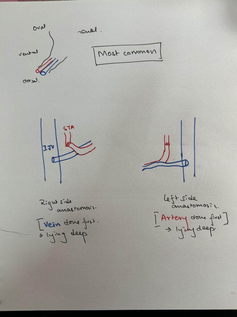
- Arteriovenous Positioning: The jejunal artery was most commonly positioned on the oral and ventral sides of the vein (48.6%).
- Surgical Significance: The artery’s position can impact microvascular anastomosis techniques, affecting surgical outcomes.
- Patient Sample: 78 patients who underwent free jejunal flap reconstruction between June 2021 and May 2023 were studied.
- Recipient Vessels: The superior thyroid artery (44.6%) and transverse cervical artery (50.0%) were the most common recipient arteries. The internal jugular vein (85.1%) was the primary recipient vein.
- Vascular Placement Patterns: Six distinct classifications (A-F) were identified based on artery and vein positioning.
- Clinical Outcome: One case of postoperative arterial thrombosis required reanastomosis but was successfully salvaged.
- Limitations: Small sample size, single-institution study, and potential positional variations due to surgical techniques.
Notes:
Optimizing Free Jejunal Flap Surgery: A Study on Arteriovenous Positioning
Free jejunal flap transfer is a crucial reconstructive technique in head and neck surgery, particularly after extensive oncologic resections such as total pharyngolaryngectomy. While the procedure is well-established, optimizing the microvascular anastomosis remains a challenge, often influenced by the intricate vascular anatomy of the jejunum. A recent study published in the Journal of Plastic, Reconstructive & Aesthetic Surgery sheds light on this issue, offering a deeper understanding of the spatial relationships between the jejunal artery and vein.
Conducted at the National Cancer Center Hospital East in Japan, this retrospective cohort study analyzed intraoperative photographs of 78 patients who underwent free jejunal flap reconstruction. The researchers classified the positioning of the jejunal artery relative to the vein into six patterns (A-F). They found that in most cases (48.6%), the artery was positioned on the oral and ventral sides of the vein, which has significant implications for surgical planning and technique.
One of the key takeaways from this study is that understanding the anatomical positioning of the jejunal vasculature can improve the efficiency of vascular anastomosis. For instance, in cases where the artery is ventrally positioned, surgeons may need to modify their approach to prevent venous compression, a common complication in microsurgery. The study also highlights that the superior thyroid and transverse cervical arteries are the most frequently used recipient vessels, while the internal jugular vein is the dominant venous choice.
However, the study has its limitations. Being a single-center study with a relatively small patient sample, its findings may not be universally applicable. Additionally, the retrospective nature of the study means that certain variables, such as intraoperative vessel manipulation, could not be fully controlled. The authors suggest that a multi-center study with a larger cohort could provide more comprehensive insights into vascular positioning patterns.
Despite these limitations, the study offers valuable guidance for plastic and reconstructive surgeons performing free jejunal flap transfers. By understanding the nuances of vascular anatomy, surgeons can improve anastomotic techniques, reduce intraoperative complications, and ultimately enhance patient outcomes.
This research adds to the growing body of literature emphasizing the importance of precise vascular mapping in reconstructive surgery. As microsurgical techniques continue to evolve, studies like this pave the way for safer and more effective surgical procedures, minimizing complications while optimizing flap viability.
Kondo, A., Higashino, T., Shimada, K., Hashimoto, K., Fukunaga, Y., Oshima, A., & Ogawa, R. (2025, February). Jejunal artery and vein positioning in free jejunal transfer: Surgical considerations and clinical implications. Journal of plastic, reconstructive & aesthetic surgery : JPRAS, 101, 84-89. https://doi.org/10.1016/j.bjps.2024.11.058